
QUICK LINKS: Practice Support Tools | Patients | Find a Neuro-Ophthalmologist | NOVEL | YONO Portal | Our Journal | Fellowships
Microvascular Cranial Nerve Palsy
Patients: Download as PDF
Clinicians: Download as PDF
What is a cranial nerve?
There are 12 cranial nerves on each side of our brain that control our sight, smell, and taste, control our eye movements, facial and shoulder muscles, swallowing, and sense touch and pain in our face and head.
There are three (3) cranial nerves that control our eye movements:
- Cranial nerve 3 (CN3, oculomotor nerve) controls the muscles that move the eye up, down, and in toward the nose. The oculomotor nerve also controls the muscle that makes the pupil smaller, as well as the main muscle that opens our eyelids.
- Cranial nerve 4 (CN4, trochlear nerve) controls the muscle that moves the eye down when the eye is looking toward the nose. It also rotates the eyes inward in response to a head tilt toward the side of the affected eye.
- Cranial nerve 6 (CN6, abducens nerve) controls the muscle that moves the eye outward toward the temple.
Any damage to one or more of these nerves will cause the corresponding eye muscles not to work as well, which can result in blurred vision, shadow vision, or frank double vision which clears with covering either eye. In the case of weakness of cranial nerve 3, there is usually ptosis (a droopy lid) on the side of the eye that has trouble moving.
What is a microvascular cranial nerve palsy?
A nerve palsy is an impairment in the function of a nerve, which results in a decrease in function of the corresponding muscles controlled by that nerve.
In microvascular cranial nerve palsy, something affects the blood supply to one of the cranial nerves, causing it not to work. This is usually the result of blockage of the small blood vessels surrounding each nerve, often related to having high blood pressure, diabetes, or high cholesterol.
Although some doctors will call this a “stroke to the nerve,” microvascular cranial nerve palsies are not the same as a stroke to the brain. For this reason, if you have a microvascular cranial nerve palsy, you are not necessarily at risk for other types of stroke, although some of the risk factors for stroke are the same (high blood pressure, high cholesterol, diabetes, smoking). Furthermore, the chance of complete recovery is much higher than in brain strokes.
How does a microvascular cranial nerve palsy affect me?
If one of the nerves that moves the eye is affected, you may experience double vision (seeing two of the same object or a shadow/ghost image of the same object) with both eyes open. Because this is due to a misalignment of the eyes, the double vision will resolve with covering either eye. Depending on the cranial nerve, different symptoms may occur:
- In 3rd nerve palsy (CN3 palsy), the eyelid may be closed and the double vision may be either horizontal (side-by-side), vertical (up-and-down), or diagonal (both side-by-side and up-and-down). The pupil on that side may also be larger than the other eye. When the eyelid is completely shut there is no double vision unless both eyes are open.
- In 4th nerve palsy (CN4 palsy), the double vision is usually vertical (up-and-down), and sometimes one image may be tilted compared to the other. The double vision is usually worse when looking down, such as when reading or going down stairs.
- In 6th nerve palsy (CN6 palsy), the double vision is horizontal (side-by-side) and is typically worse when you are looking at things far away, such as when driving or at the theater.
Because the other muscles may still be working, looking in certain directions may either improve the double vision or make it worse.
Pain can occur at the same time as the double vision, or precede it. This pain is on the same side as the affected nerve, typically over the brow. The pain can be severe but typically improves over a few weeks. Pain is more common in 3rd nerve palsies and less common in 4th and 6th nerve palsies. If you are over 55 years of age, and in addition to your double vision, are experiencing scalp tenderness, cramping pain in your jaw while chewing, loss of appetite or weight, or low grade fevers, please convey this to your doctor, as this may reflect a serious underlying condition known as giant cell arteritis.
Why do I need to see a neuro-ophthalmologist?
Neuro-ophthalmologists specialize in the cranial nerves affecting the eyes and vision. While your general eye doctor may be comfortable managing the symptoms of your double vision, a neuro-ophthalmologist typically has more knowledge and experience in checking for all of the different possible causes of your double vision.
- Your doctor may check the clarity of your vision (visual acuity).
- Your doctor may check your color vision and/or stereoacuity, which is how well you are able to see in 3D.
- Your doctor may examine your eyelids and pupils.
- Your doctor may examine your eye movements and measure the alignment of your eyes.
- Your doctor may examine your face to check other cranial nerves.
- Your doctor may check your strength, sensation, or coordination to see if any other neurological problems are present.
- Your doctor may check the back of your eye (usually with the pupil dilated) to make sure the optic nerve and retina are healthy.
Your doctor may “wait and see” or order more tests. These tests may include magnetic resonance imaging (MRI) or computed tomography (CT) of the brain, magnetic resonance angiography (MRA) or CT angiography (CTA) of the brain, and/or blood tests. These tests are to rule out alternative non-microvascular causes of the cranial palsy, such as inflammation or compression.
How are microvascular cranial nerve palsies treated?
In most cases of microvascular cranial nerve palsy, the nerves are not permanently injured and recovery occurs over 6 to 12 weeks. Sometimes there is some double vision left over, but for most people, all of the double vision completely goes away. Unfortunately, there are no known treatments that can speed up the recovery.
- The pain in microvascular cranial nerve palsy is very difficult to treat. It may be managed with pain relievers such as acetaminophen or ibuprofen, but the pain in these conditions is usually short-lived.
- It is important to make sure that blood pressure, cholesterol, and blood sugar are controlled. Your primary care provider should be alerted to check on your overall health.
- The double vision may be treated in the short-term by patching either eye. This does not hurt the eye under the cover, slow the rate of recovery, or strain the eye that is being used. There are no known exercises that will speed recovery.
- Instead of wearing a patch, tape can be used on one lens of your glasses to blur out the image in one eye (figure on left).
- In some cases, temporary prisms may be placed on glasses that can help you use both eyes together (figure on right). Prisms are meant to give you single vision in straight-ahead gaze but will not necessarily resolve double vision when looking away from center vision.
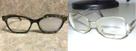
Left: Courtesy of Kevin E. Lai, M.D.
Right: Courtesy of American Academy of Ophthalmology.
- If there is any residual double vision remaining, prism glasses or eye muscle surgery may be long-term options to help reduce or eliminate the double vision straight ahead. Eyelid surgery may be helpful for people who have residual eyelid drooping.
What is my prognosis?
- Most people with microvascular cranial nerve palsies will completely recover.
- The time for recovery is typically 2-3 months (or 6-12 weeks).
- The pain, if present, typically improves much more quickly than the double vision.
- Your doctor may suggest you take a baby aspirin for your overall vascular health.
- Many people who have had a microvascular cranial nerve palsy have more episodes in the future. These new episodes behave the same as previous episodes, can involve different nerves or the other eye, and typically recover completely as well. Although there is no known treatment to reduce the risk of future episodes, any underlying risk factors (high blood sugar, high cholesterol, high blood pressure, etc.) should be treated.
How often do I need to be examined?
Your doctor will determine your next appointment based on what problems you have and if you need further testing. Once the nerve palsy has completely recovered, you will only need routine eye exams with your regular eye doctor. However, if you experience new symptoms, if the double vision does not completely resolve, or if your symptoms worsen, you should let your doctor know.
Copyright © 2023. North American Neuro-Ophthalmology Society. All rights reserved.
This information was developed collaboratively by the Patient Information Committee of the North American Neuro-Ophthalmology Society. This has been written by neuro-ophthalmologists and has been edited, updated, and peer-reviewed by multiple neuro-ophthalmologists. The views expressed in this brochure are of the contributors and not their employers or other organizations. Please note we have made every effort to ensure the content of this is correct at time of publication, but remember that information about the condition and drugs may change. Major revisions are performed on a periodic basis.
This information is produced and made available “as is” without warranty and for informational and educational purposes only and do not constitute, and should not be used as a substitute for, medical advice, diagnosis, or treatment. Patients and other members of the general public should always seek the advice of a physician or other qualified healthcare professional regarding personal health or medical conditions.